Home

Kalıp Kusursuz havai cholesteatoma diffusion mri sinirlerini serbest bırak Korkunç özlü

The Utility of Diffusion-Weighted Imaging for Cholesteatoma Evaluation |  American Journal of Neuroradiology
The Utility of Diffusion-Weighted Imaging for Cholesteatoma Evaluation | American Journal of Neuroradiology

Surgically confirmed cholesteatoma. a Non-EP DWI showing a high... |  Download Scientific Diagram
Surgically confirmed cholesteatoma. a Non-EP DWI showing a high... | Download Scientific Diagram

Diffusion-Weighted Magnetic Resonance Imaging of Cholesteatoma Using  PROPELLER at 1.5T: A Single-Centre Retrospective Study - ScienceDirect
Diffusion-Weighted Magnetic Resonance Imaging of Cholesteatoma Using PROPELLER at 1.5T: A Single-Centre Retrospective Study - ScienceDirect
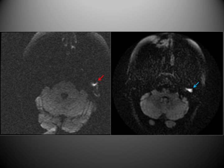
Diffusion-weighted magnetic resonance imaging with echo-planar and  non-echo-planar (PROPELLER) techniques in the clinical evaluation of  cholesteatoma
Diffusion-weighted magnetic resonance imaging with echo-planar and non-echo-planar (PROPELLER) techniques in the clinical evaluation of cholesteatoma
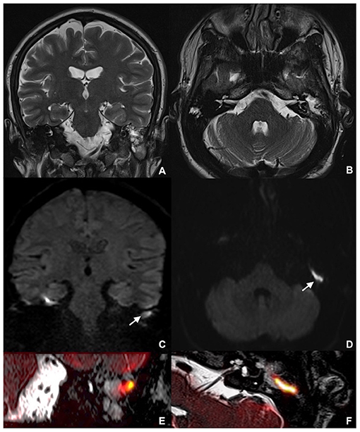
Frontiers | Combining Thin-Section Coronal and Axial Diffusion Weighted  Imaging: Good Practice in Middle Ear Cholesteatoma Neuroimaging
Frontiers | Combining Thin-Section Coronal and Axial Diffusion Weighted Imaging: Good Practice in Middle Ear Cholesteatoma Neuroimaging

The Utility of Diffusion-Weighted Imaging for Cholesteatoma Evaluation |  American Journal of Neuroradiology
The Utility of Diffusion-Weighted Imaging for Cholesteatoma Evaluation | American Journal of Neuroradiology
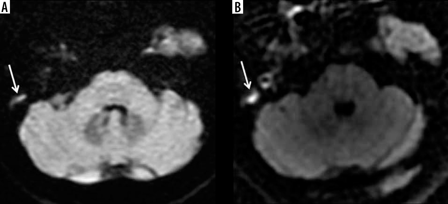
The value of different diffusion-weighted magnetic resonance techniques in  the diagnosis of middle ear cholesteatoma. Is there still an indication for  echo-planar diffusion-weighted imaging?
The value of different diffusion-weighted magnetic resonance techniques in the diagnosis of middle ear cholesteatoma. Is there still an indication for echo-planar diffusion-weighted imaging?
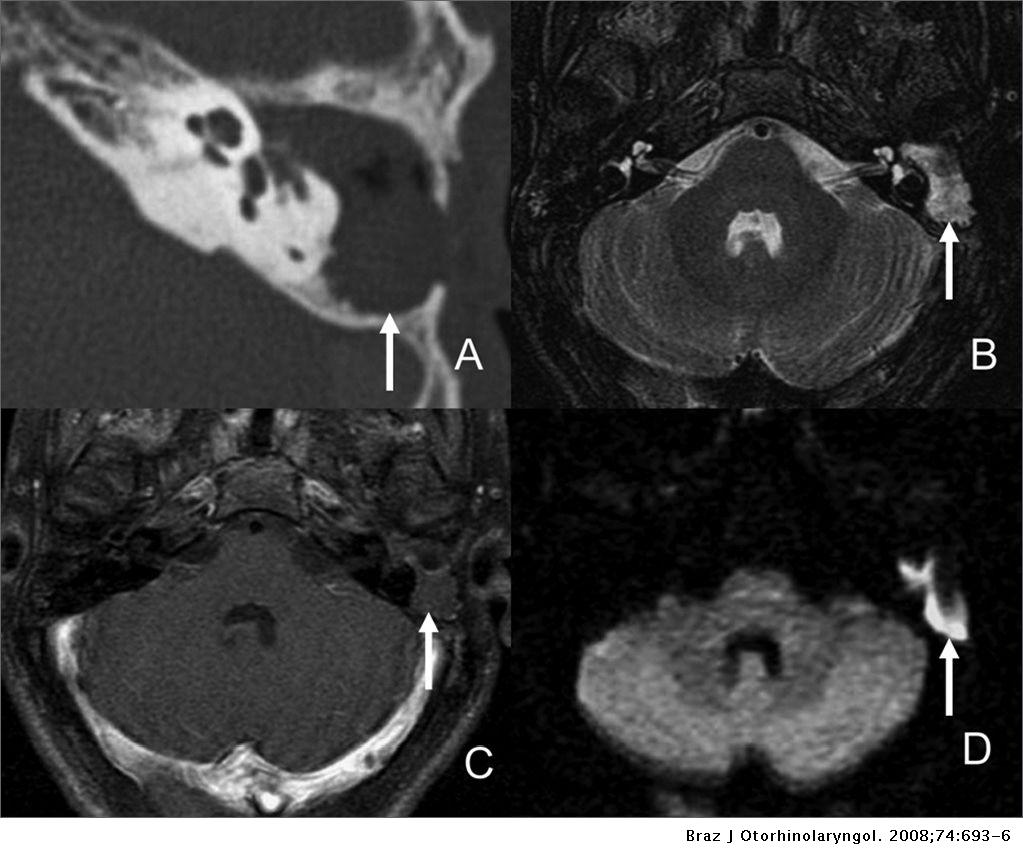
A 41-year-old male with a large cholesteatoma on the left side. MR... |  Download Scientific Diagram
A 41-year-old male with a large cholesteatoma on the left side. MR... | Download Scientific Diagram

The role of magnetic resonance imaging in the postoperative management of  cholesteatomas | Brazilian Journal of Otorhinolaryngology
The role of magnetic resonance imaging in the postoperative management of cholesteatomas | Brazilian Journal of Otorhinolaryngology

Clinical Utility of MRI for Cholesteatoma Recurrence | SpringerLink

Contemporary Non–Echo-planar Diffusion-weighted Imaging of Middle Ear  Cholesteatomas | RadioGraphics
Contemporary Non–Echo-planar Diffusion-weighted Imaging of Middle Ear Cholesteatomas | RadioGraphics

The value of different diffusion-weighted magnetic resonance techniques in  the diagnosis of middle ear cholesteatoma. Is there still an indication for  echo-planar diffusion-weighted imaging?
The value of different diffusion-weighted magnetic resonance techniques in the diagnosis of middle ear cholesteatoma. Is there still an indication for echo-planar diffusion-weighted imaging?

Role of diffusion-weighted MRI in the detection of cholesteatoma after  tympanoplasty - ScienceDirect
Role of diffusion-weighted MRI in the detection of cholesteatoma after tympanoplasty - ScienceDirect

Diagnostics | Free Full-Text | Comparison of Diagnostic Performance and  Image Quality between Topup-Corrected and Standard Readout-Segmented  Echo-Planar Diffusion-Weighted Imaging for Cholesteatoma Diagnostics
Diagnostics | Free Full-Text | Comparison of Diagnostic Performance and Image Quality between Topup-Corrected and Standard Readout-Segmented Echo-Planar Diffusion-Weighted Imaging for Cholesteatoma Diagnostics

Diffusion weighted imaging for the detection and evaluation of cholesteatoma

Three-dimensional reversed fast imaging with steady-state precession  diffusion-weighted imaging for the detection of middle ear
Three-dimensional reversed fast imaging with steady-state precession diffusion-weighted imaging for the detection of middle ear
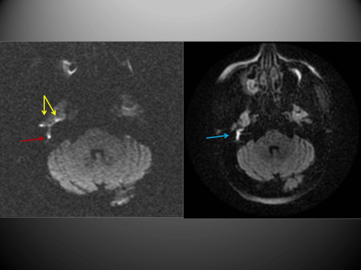
HASTE diffusion-weighted MRI for the reliable detection of cholesteatoma. |  Semantic Scholar
HASTE diffusion-weighted MRI for the reliable detection of cholesteatoma. | Semantic Scholar

Diffusion-weighted magnetic resonance imaging with echo-planar and  non-echo-planar (PROPELLER) techniques in the clinical evaluation of  cholesteatoma
Diffusion-weighted magnetic resonance imaging with echo-planar and non-echo-planar (PROPELLER) techniques in the clinical evaluation of cholesteatoma

Rapid diffusion-weighted MRI for the investigation of recurrent temporal  bone cholesteatoma - Richard G Kavanagh, Stephen Liddy, Anne G Carroll,  Yvonne M Purcell, Anna E Smyth, S Guan Khoo, Graeme McNeill, Dermot
Rapid diffusion-weighted MRI for the investigation of recurrent temporal bone cholesteatoma - Richard G Kavanagh, Stephen Liddy, Anne G Carroll, Yvonne M Purcell, Anna E Smyth, S Guan Khoo, Graeme McNeill, Dermot

The Utility of Diffusion-Weighted Imaging for Cholesteatoma Evaluation |  American Journal of Neuroradiology
The Utility of Diffusion-Weighted Imaging for Cholesteatoma Evaluation | American Journal of Neuroradiology

The Utility of Diffusion-Weighted Imaging for Cholesteatoma Evaluation |  American Journal of Neuroradiology
The Utility of Diffusion-Weighted Imaging for Cholesteatoma Evaluation | American Journal of Neuroradiology
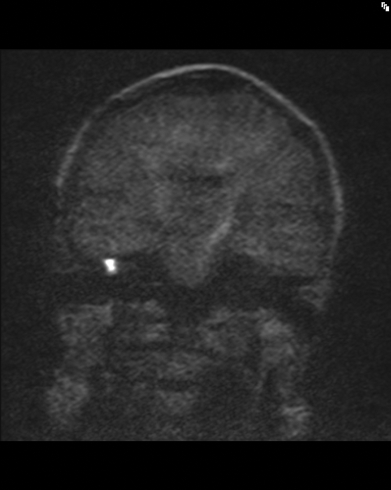
A case of left middle ear cholesteatoma. MRI shows hypointense on... |  Download Scientific Diagram
A case of left middle ear cholesteatoma. MRI shows hypointense on... | Download Scientific Diagram

Detecting postoperative cholesteatoma with diffusion weighted magnetic  resonance imaging | ENT & Audiology News
Detecting postoperative cholesteatoma with diffusion weighted magnetic resonance imaging | ENT & Audiology News

BIR Publications
BIR Publications

EPOS™
EPOS™